The human eye gives us more information about the outside world than any other sensory organ, producing continuous images that are instantly transmitted to the brain for processing. Not only is the eye a personal window on the world, but it offers a noninvasive and immediate view into the body’s vascular system, providing physicians with an opportunity for early diagnosis of hypertension and diabetes.
In an adult, the eye has a diameter of approximately 25mm (or one inch). It sits in a cavity in the skull called the eye’s orbit. The one sixth of the eye’s surface that is exposed beyond the orbit is protected against strong light and foreign objects by the eyelids, eyelashes, and eyebrows.
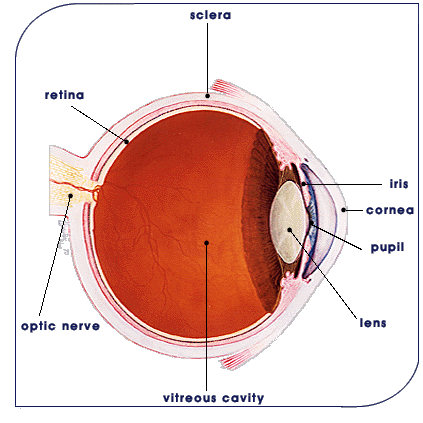
The outermost layer of the eyeball is the visible white of the eye, the sclera, which provides structure and strength; it is covered by a thin membrane called the conjunctiva.
The clear substance located inside the sclera is called the vitreous, a gel-like material that gives the eye its spherical shape. In front of the sclera is the transparent, protective cornea, which provides most of the focusing power for light entering the eye. The cornea’s outermost layer of tissue contains cells that have the ability to regenerate within three days, allowing for rapid healing of superficial injuries. From the cornea, light passes through the pupil, the dark circle centered in the iris, the blue, green, brown or hazel ring of color that helps describe a person’s appearance. The eye’s iris also functions like the iris of a camera, opening and closing to regulate the pupil’s size, which controls how much light penetrates the eye, by becoming smaller under bright conditions, or expanding in a dim environment.
Behind the iris, the lens provides fine-tuning for focusing and reading by altering its shape. The lens directs light onto the fine nerve tissue of the retina, which lines the inside wall of the eye and acts like the film in a camera. The retina converts the light into images and then into electrical impulses that are sent along the optic nerve to the brain. Within the brain, these signals undergo processing by the visual cortex, which senses and interprets them as the shapes and colors that the eye sees.
![]()
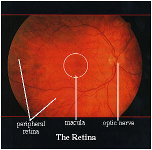
Anatomy of the Retina
The natural lens of the eye is located behind the colored part of the eye known as the iris. The lens is like a magnifying glass. The lens is transparent and helps to focus light rays onto the retina in the back of the eye.
The retina has two parts: the peripheral retina and the macula. If you imagine the retina as a circle with a bull’s-eye at the center, the macula is like the bull’s-eye, it is very small. It is located near the optic nerve. The large area of retina that surrounds the macula and makes up 95% of the retina is called the peripheral retina.
The peripheral retina gives us vision to the side, this is called “peripheral” vision. This is what we refer to when we say, “I saw something out of the corner of my eye”. Because the peripheral retina cannot see detail clearly, we cannot use peripheral vision to read, thread a needle, drive, or even recognize a face. If I see someone off to my side, “out of the corner of my eye”, I can tell who the person is by his or her general shape, but I won’t be able to see the expression on that person’s face.
In order to see fine detail, we must look straight ahead, using the macula, the bull’s-eye center of the retina. Though the macula makes up only a small part of the retina, it is one hundred times more sensitive to detail than the peripheral retina. The macula allows us to see tiny detail, to read fine print, recognize faces, thread a needle, read the time, see street signs, see grains of salt being poured from a shaker, etc.
![]()