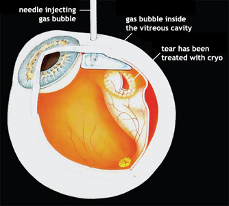
Another type of surgery that can be done for some retinal detachments is called pneumatic retinopexy. Pneumatic retinopexy is performed in the office using local anesthesia.
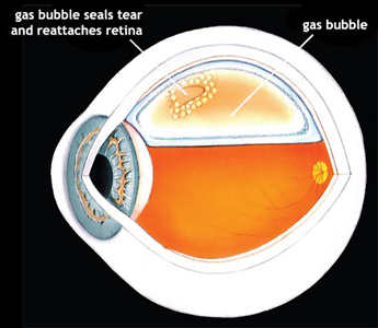
Cryotherapy or laser treatment is performed to seal the retinal tear. Instead of placing a scleral buckle on the outside of the eye, using a needle, the surgeon injects a gas bubble inside the vitreous cavity of the eye. The patient is instructed to keep his or her head in a specific position so that the gas bubble pushes the detached retina against the back wall of the eye to seal the retinal tear. The patient is asked to remain in this position for a period of time until the retinal tear is sealed against the back wall of the eye. Your surgeon will tell you how long special positioning is necessary.
The gas bubble in the vitreous cavity of the eye expands for several days and takes two to six weeks to disappear. During this time, airplane travel or travel to a high altitude must be avoided because high altitudes can result in an expansion of gas and an increase in pressure that can damage the eye. Your surgeon will tell you when it is safe to travel.
It is also important for a patient with a gas bubble not to lie face up, as the gas bubble will come to rest against the lens of the eye and may cause a cataract or high pressure in the eye. Antibiotic eye drops may be used during the days following the surgery.
The chance of successfully reattaching the retina with pneumatic retinopexy is slightly less than with a vitrectomy or scleral buckle . But with pneumatic retinopexy, hospitalization, general anesthesia, and the cutting that is necessary with scleral buckling surgery are all avoided. Pneumatic retinopexy cannot be used for every retinal detachment. Your surgeon will discuss with you whether pneumatic retinopexy is feasible and the chances for successfully reattaching your retina. Complications of pneumatic retinopexy include cataract formation, glaucoma, gas getting under the retina, excessive scar tissue formation, and infection. Any one of these complications can lead to a total loss of vision, but each is rare. The most common complication is the formation of new retinal tears and recurrence of the retinal detachment. If the retina becomes detached again, scleral buckling surgery or vitrectomy can usually be performed to reattach it.
![]()